The Orbicularis oculi (Orbicularis palpebrarum) (Fig. 379)arises from the nasal part of the frontal bone, from the frontal process of the maxilla in front of the lacrimal groove, and from the anterior surface and borders of a short fibrous band, the medial palpebral ligament. From this origin, the fibers are directed lateralward, forming a broad and thin layer, which occupies the eyelids or palpebræ, surrounds the circumference of the orbit, and spreads over the temple, and downward on the cheek. The palpebral portion of the muscle is thin and pale; it arises from the bifurcation of the medial palpebral ligament, forms a series of concentric curves, and is inserted into the lateral palpebral raphé. The orbital portion is thicker and of a reddish color; its fibers form a complete ellipse without interruption at the lateral palpebral commissure; the upper fibers of this portion blend with the Frontalis and Corrugator. The lacrimal part (Tensor tarsi) is a small, thin muscle, about 6 mm. in breadth and 12 mm. in length, situated behind the medial palpebral ligament and lacrimal sac (Fig. 379). It arises from the posterior crest and adjacent part of the orbital surface of the lacrimal bone, and passing behind the lacrimal sac, divides into two slips, upper and lower, which are inserted into the superior and inferior tarsi medial to the puncta lacrimalia; occasionally it is very indistinct.
The medial palpebral ligament (tendo oculi), about 4 mm. in length and 2 mm. in breadth, is attached to the frontal process of the maxilla in front of the lacrimal groove. Crossing the lacrimal sac, it divides into two parts, upper and lower, each attached to the medial end of the corresponding tarsus. As the ligament crosses the lacrimal sac, a strong aponeurotic lamina is given off from its posterior surface; this expands over the sac, and is attached to the posterior lacrimal crest.
The lateral palpebral raphé is a much weaker structure than the medial palpebral ligament. It is attached to the margin of the frontosphenoidal process of the zygomatic bone, and passes medialward to the lateral commissure of the eyelids, where it divides into two slips, which are attached to the margins of the respective tarsi.
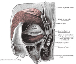
FIG. 379– Left orbicularis oculi, seen from behind. (See enlarged image)
The Corrugator79 (Corrugator supercilii) is a small, narrow, pyramidal muscle, placed at the medial end of the eyebrow, beneath the Frontalis and Orbicularis oculi. It arises from the medial end of the superciliary arch; and its fibers pass upward and lateralward, between the palpebral and orbital portions of the Orbicularis oculi, and are inserted into the deep surface of the skin, above the middle of the orbital arch.
Actions.—The Orbicularis oculi is the sphincter muscle of the eyelids. The palpebral portion acts involuntarily, closing the lids gently, as in sleep or in blinking; the orbital portion is subject to the will. When the entire muscle is brought into action, the skin of the forehead, temple, and cheek is drawn toward the medial angle of the orbit, and the eyelids are firmly closed, as in photophobia. The skin thus drawn upon is thrown into folds, especially radiating from the lateral angle of the eyelids; these folds become permanent in old age, and form the so-called “crows’ feet.” The Levator palpebræ superioris is the direct antagonist of this muscle; it raises the upper eyelid and exposes the front of the bulb of the eye. Each time the eyelids are closed through the action of the Orbicularis, the medial palpebral ligament is tightened, the wall of the lacrimal sac is thus drawn lateralward and forward, so that a vacuum is made in it and the tears are sucked along the lacrimal canals into it. The lacrimal part of the Orbicularis oculi draws the eyelids and the ends of the lacrimal canals medialward and compresses them against the surface of the globe of the eye, thus placing them in the most favorable situation for receiving the tears; it also compresses the lacrimal sac. The Corrugator draws the eyebrow downward and medialward, producing the vertical wrinkles of the forehead. It is the “frowning” muscle, and may be regarded as the principal muscle in the expression of suffering.
The United States Medical Licensing Examination (USMLE) is a three-step examination for medical licensure in the United States. The Federation of State Medical Boards (FSMB) and the National Board of Medical Examiners (NBME) sponsors USMLE.
The Three Steps of the USMLE
Step 1 tests the important concepts of basic sciences basic to the practice of medicine. It also places special emphasis on principles and mechanisms underlying health, disease, and modes of therapy. Step 1 ensures mastery of the sciences that provide a foundation for the safe and competent practice of medicine. It also tests the scientific principles required for maintenance of competence through lifelong learning.
Step 2 CK tests the medical knowledge, skills, and understanding of clinical science essential for the provision of patient care under supervision. It also includes emphasis on health promotion and disease prevention. Step 2 CK ensures that due attention is devoted to principles of clinical sciences and basic patient-centered skills.
Step 2 CS tests your capacity to practice and provide good medical service in real-life situations. It also tests your communication skills.
Step 3 tests your medical knowledge and understanding of biomedical and clinical science essential for the unsupervised practice of medicine. Step 3 provides a final assessment of physicians assuming independent responsibility for delivering general medical care.