5b. Articulation of the Atlas with the Epistropheus or Axis
(Articulatio Atlantoepistrophica)
The articulation of the atlas with the axis is of a complicated nature, comprising no fewer than four distinct joints. There is a pivot articulation between the odontoid process of the axis and the ring formed by the anterior arch and the tranverse ligament of the atlas (see Fig. 306); here there are two joints: one between the posterior surface of the anterior arch of the atlas and the front of the odontoid process; the other between the anterior surface of the ligament and the back of the process. Between the articular processes of the two bones there is on either side an arthrodial or gliding joint. The ligaments connecting these bones are:
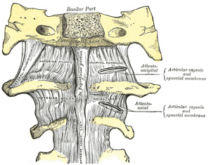
FIG. 304– Anterior atlantoöccipital membrane and atlantoaxial ligament. (See enlarged image)
The Articular Capsules (capsulæ articulares; capsular ligaments).—The articular capsules are thin and loose, and connect the margins of the lateral masses of the atlas with those of the posterior articular surfaces of the axis. Each is strengthened at its posterior and medial part by an accessory ligament, which is attached below to the body of the axis near the base of the odontoid process, and above to the lateral mass of the atlas near the transverse ligament.
The Anterior Atlantoaxial Ligament (Fig. 304).—This ligament is a strong membrane, fixed, above, to the lower border of the anterior arch of the atlas; below, to the front of the body of the axis. It is strengthened in the middle line by a rounded cord, which connects the tubercle on the anterior arch of the atlas to the body of the axis, and is a continuation upward of the anterior longitudinal ligament. The ligament is in relation, in front, with the Longi capitis.
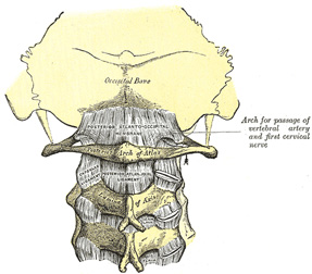
FIG. 305– Posterior atlantoöccipital membrane and atlantoaxial ligament. (See enlarged image)
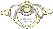
FIG. 306– Articulation between odontoid process and atlas. (See enlarged image)
The Posterior Atlantoaxial Ligament (Fig. 305).—This ligament is a broad, thin membrane attached, above, to the lower border of the posterior arch of the atlas; below, to the upper edges of the laminæ of the axis. It supplies the place of the ligamenta flava, and is in relation, behind, with the Obliqui capitis inferiores.
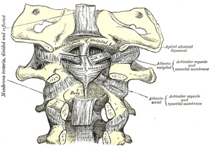
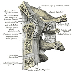
FIG. 307– Membrana tectoria, transverse, and alar ligaments. (See enlarged image)
FIG. 308– Median sagittal section through the occipital bone and first three cervical vertebræ. (Spalteholz.) (See enlarged image)
The Transverse Ligament of the Atlas (ligamentum transversum atlantis) (Fig. 306, 307, 308).—The transverse ligament of the atlas is a thick, strong band, which arches across the ring of the atlas, and retains the odontoid process in contact with the anterior arch. It is concave in front, convex behind, broader and thicker in the middle than at the ends, and firmly attached on either side to a small tubercle on the medial surface of the lateral mass of the atlas. As it crosses the odontoid process, a small fasciculus (crus superius) is prolonged upward, and another (crus inferius) downward, from the superficial or posterior fibers of the ligament. The former is attached to the basilar part of the occipital bone, in close relation with the membrana tectoria; the latter is fixed to the posterior surface of the body of the axis; hence, the whole ligament is named the cruciate ligament of the atlas. The transverse ligament divides the ring of the atlas into two unequal parts: of these, the posterior and larger serves for the transmission of the medulla spinalis and its membranes and the accessory nerves; the anterior and smaller contains the odontoid process. The neck of the odontoid process is constricted where it is embraced posteriorly by the transverse ligament, so that this ligament suffices to retain the odontoid process in position after all the other ligaments have been divided.
Synovial Membranes.—There is a synovial membrane for each of the four joints; the joint cavity between the odontoid process and the transverse ligament is often continuous with those of the atlantoöccipital articulations.
Movements.—The opposed articular surfaces of the atlas and axis are not reciprocally curved; both surfaces are convex in their long axes. When, therefore, the upper facet glides forward on the lower it also descends; the fibers of the articular capsule are relaxed in a vertical direction, and will then permit of movement in an antero-posterior direction. By this means a shorter capsule suffices and the strength of the joint is materially increased.67
The principal muscles by which these movements are produced are the Sternocleidomastoideus and Semispinalis capitis of one side, acting with the Longus capitis, Splenius, Longissimus capitis, Rectus capitis posterior major, and Obliqui capitis superior and inferior of the other side.
Note 67. Corner (“The Physiology of the Atlanto-axial Joints,” Journal of Anatomy and Physiology, vol. xli) states that the movements which take place at these articulations are of a complex nature. The first part of the movement is an eccentric or asymmetrical one; the atlanto-axial joint of the side to which the head is moved is fixed, or practically fixed, by the muscles of the neck, and forms the center of the movement, while the opposite atlantal facet is carried downward and forward on the corresponding axial facet. The second part of the movement is centric and symmetrical, the odontoid process forming the axis of the movement [back]
You are welcome to ask for hospital review for residency. We will be providing them to those who ask them first.
The United States Medical Licensing Examination (USMLE) is a three-step examination for medical licensure in the United States. The Federation of State Medical Boards (FSMB) and the National Board of Medical Examiners (NBME) sponsors USMLE.
The Three Steps of the USMLE
Step 1 tests the important concepts of basic sciences basic to the practice of medicine. It also places special emphasis on principles and mechanisms underlying health, disease, and modes of therapy. Step 1 ensures mastery of the sciences that provide a foundation for the safe and competent practice of medicine. It also tests the scientific principles required for maintenance of competence through lifelong learning.
Step 2 CK tests the medical knowledge, skills, and understanding of clinical science essential for the provision of patient care under supervision. It also includes emphasis on health promotion and disease prevention. Step 2 CK ensures that due attention is devoted to principles of clinical sciences and basic patient-centered skills.
Step 2 CS tests your capacity to practice and provide good medical service in real-life situations. It also tests your communication skills.
Step 3 tests your medical knowledge and understanding of biomedical and clinical science essential for the unsupervised practice of medicine. Step 3 provides a final assessment of physicians assuming independent responsibility for delivering general medical care.