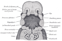
FIG. 947– The head and neck of a human embryo thirty-two days old, seen from the ventral surface. The floor of the mouth and pharynx have been removed. (His.) (See enlarged image)
Development.—The rudiment of the respiratory organs appears as a median longitudinal groove in the ventral wall of the pharynx. The groove deepens and its lips fuse to form a septum which grows from below upward and converts the groove into a tube, the laryngo-tracheal tube(Fig. 947), the cephalic end of which opens into the pharynx by a slit-like aperture formed by the persistent anterior part of the groove. The tube is lined by entoderm from which the epithelial lining of the respiratory tract is developed. The cephalic part of the tube becomes the larynx, and its next succeeding part the trachea, while from its caudal end two lateral outgrowths, the right and left lung buds, arise, and from them the bronchi and lungs are developed. The first rudiment of the larynx consists of two arytenoid swellings, which appear, one on either side of the cephalic end of the laryngo-tracheal groove, and are continuous in front of the groove with a transverse ridge (furcula of His) which lies between the ventral ends of the third branchial arches and from which the epiglottis is subsequently developed (Figs. 980,981). After the separation of the trachea from the esophagus the arytenoid swellings come into contact with one another and with the back of the epiglottis, and the entrance to the larynx assumes the form of a T-shaped cleft, the margins of the cleft adhere to one another and the laryngeal entrance is for a time occluded. The mesodermal wall of the tube becomes condensed to form the cartilages of the larynx and trachea. The arytenoid swellings are differentiated into the arytenoid and corniculate cartilages, and the folds joining them to the epiglottis form the aryepiglottic folds in which the cuneiform cartilages are developed as derivatives of the epiglottis. The thyroid cartilage appears as two lateral plates, each chondrified from two centers and united in the mid-ventral line by membrane in which an additional center of chondrification develops. The cricoid cartilage arises from two cartilaginous centers, which soon unite ventrally and gradually extend and ultimately fuse on the dorsal aspect of the tube.
The opening of the pulmonary diverticulum lies between the two fifth arch masses and behind a “central mass” in the middle line—the proximal end of the diverticulum is compressed between the fifth arch masses. The fifth arch is joined by the fourth to form a “lateral mass” on each side of the opening, and these “lateral masses” grow forward and overlap the central mass and so form a secondary transverse cavity, which is really a part of the cavity of the pharynx. The two parts of the cavity of the larynx are separated in the adult by a line drawn back along the vocal fold and then upward along the border of the arytenoid eminence to the interarytenoid notch. The arytenoid and cricoid are developed in the fifth arch mass. The thyroid is primarily a fourth arch derivative, and if it has a fifth arch element this is a later addition. The epiglottis is derived from the “central mass,” and has a third arch element in its oral and upper aspect; the arch value of the “central mass” is doubtful.
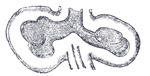
FIG. 948– Lung buds from a human embryo of about four weeks, showing commencing lobulations. (His.) (See enlarged image)
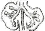
FIG. 949– Lungs of a human embryo more advanced in development. (His.) (See enlarged image)
The right and left lung buds grow out behind the ducts of Cuvier, and are at first symmetrical, but their ends soon become lobulated, three lobules appearing on the right, and two on the left; these subdivisions are the early indications of the corresponding lobes of the lungs (Figs. 948,949). The buds undergo further subdivision and ramification, and ultimately end in minute expanded extremities—the infundibula of the lung. After the sixth month the air-sacs begin to make their appearance on the infundibula in the form of minute pouches. The pulmonary arteries are derived from the sixth aortic arches. During the course of their development the lungs migrate in a caudal direction, so that by the time of birth the bifurcation of the trachea is opposite the fourth thoracic vertebra. As the lungs grow they project into that part of the celom which will ultimately form the pleural cavities, and the superficial layer of the mesoderm enveloping the lung rudiment expands on the growing lung and is converted into the pulmonary pleura.
The United States Medical Licensing Examination (USMLE) is a three-step examination for medical licensure in the United States. The Federation of State Medical Boards (FSMB) and the National Board of Medical Examiners (NBME) sponsors USMLE.
The Three Steps of the USMLE
Step 1 tests the important concepts of basic sciences basic to the practice of medicine. It also places special emphasis on principles and mechanisms underlying health, disease, and modes of therapy. Step 1 ensures mastery of the sciences that provide a foundation for the safe and competent practice of medicine. It also tests the scientific principles required for maintenance of competence through lifelong learning.
Step 2 CK tests the medical knowledge, skills, and understanding of clinical science essential for the provision of patient care under supervision. It also includes emphasis on health promotion and disease prevention. Step 2 CK ensures that due attention is devoted to principles of clinical sciences and basic patient-centered skills.
Step 2 CS tests your capacity to practice and provide good medical service in real-life situations. It also tests your communication skills.
Step 3 tests your medical knowledge and understanding of biomedical and clinical science essential for the unsupervised practice of medicine. Step 3 provides a final assessment of physicians assuming independent responsibility for delivering general medical care.