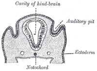
FIG. 898– Section through the head of a human embryo, about twelve days old, in the region of the hind-brain. (Kollmann.) (See enlarged image)
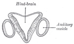
FIG. 899– Section through hind-brain and auditory vesicles of an embryo more advanced than that of Fig. 898. (After His.) (See enlarged image)
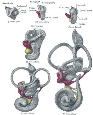
FIG. 900– Lateral views of membranous labyrinth and acoustic complex. X 25 dia. (Streeter.) absorpt. focu, area of wall where absorption is complete; amp., ampulla membranacea; crus, crus commune; d. sc. lat., ductus semicircularis lateralis; d. sc. post., ductus semicircularis posterior; d. sc. sup., ductus semicircular superior; coch. or cochlea, ductus cochlearis; duct. endolymph, ductus endolymphaticus; d. reuniens, ductus reuniens Henseni; endol. or endolymphs appendix endolymphaticus; rec. utr., recessus utriculi; sacc., sacculus; sac. endol., saccus endolymphaticus; sinus utr. lat., sinus utriculi lateralis; utric., utriculus; vestib. p., vestibular pouch. (See enlarged image)
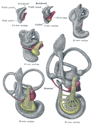
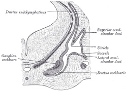
The Development of the Ear.—The first rudiment of the internal ear appears shortly after that of the eye, in the form of a patch of thickened ectoderm, the auditory plate, over the region of the hind-brain. The auditory plate becomes depressed and converted into the auditory pit(Fig. 898). The mouth of the pit is then closed, and thus a shut sac, the auditory vesicle, is formed (Fig. 899); from it the epithelial lining of the membranous labyrinth is derived. The vesicle becomes pear-shaped, and the neck of the flask is obliterated (Fig. 900). From the vesicle certain diverticula are given off which form the various parts of the membranous labyrinth. One from the middle part forms the ductus and saccus endolymphaticus, another from the anterior end gradually elongates, and, forming a tube coiled on itself, becomes the cochlear duct, the vestibular extremity of which is subsequently constricted to form the canalis reuniens. Three others appear as disk-like evaginations on the surface of the vesicle; the central parts of the walls of the disks coalesce and disappear, while the peripheral portions persist to form the semicircular ducts; of these the superior is the first and the lateral the last to be completed (Fig. 902). The central part of the vesicle represents the membranous vestibule, and is subdivided by a constriction into a smaller ventral part, the saccule, and a larger dorsal and posterior part, the utricle. This subdivision is effected by a fold which extends deeply into the proximal part of the ductus endolymphaticus, with the result that the utricle and saccule ultimately communicate with each other by means of a Y-shaped canal. The saccule opens into the cochlear duct, through the canalis reuniens, and the semicircular ducts communicate with the utricle.
FIG. 901– Median views of membranous labyrinth and acoustic complex in human embryos. X 25 dia. (Streeter.) (See enlarged image)
FIG. 902– Transverse section through head of fetal sheep, in the region of the labyrinth. X 30. (After Boettcher.) (See enlarged image)
The mesodermal tissue surrounding the various parts of the epithelial labyrinth is converted into a cartilaginous ear-capsule, and this is finally ossified to form the bony labyrinth. Between the cartilaginous capsule and the epithelial structures is a stratum of mesodermal tissue which is differentiated into three layers, viz., an outer, forming the periosteal lining of the bony labyrinth; an inner, in direct contact with the epithelial structures; and an intermediate, consisting of gelatinous tissue: by the absorption of this latter tissue the perilymphatic spaces are developed. The modiolus and osseous spiral lamina of the cochlea are not preformed in cartilage but are ossified directly from connective tissue.
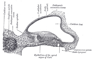
FIG. 903– Transverse section of the cochlear duct of a fetal cat. (After Boettcher and Ayres.) (See enlarged image)
The middle ear and auditory tube are developed from the first pharyngeal pouch. The entodermal lining of the dorsal end of this pouch is in contact with the ectoderm of the corresponding pharyngeal groove; by the extension of the mesoderm between these two layers the tympanic membrane is formed. During the sixth or seventh month the tympanic antrum appears as an upward and backward expansion of the tympanic cavity. With regard to the exact mode of development of the ossicles of the middle ear there is some difference of opinion. The view generally held is that the malleus is developed from the proximal end of the mandibular (Meckel’s) cartilage (Fig. 43), the incus in the proximal end of the mandibular arch, and that the stapes is formed from the proximal end of the hyoid arch. The malleus, with the exception of its anterior process is ossified from a single center which appears near the neck of the bone; the anterior process is ossified separately in membrane and joins the main part of the bone about the sixth month of fetal life. The incus is ossified from one center which appears in the upper part of its long crus and ultimately extends into its lenticular process. The stapes first appears as a ring (annulus stapedius) encircling a small vessel, the stapedial artery, which subsequently undergoes atrophy; it is ossified from a single center which appears in its base.
The external acoustic meatus is developed from the first branchial groove. The lower part of this groove extends inward as a funnel-shaped tube (primary meatus) from which the cartilaginous portion and a small part of the roof of the osseous portion of the meatus are developed. From the lower part of the funnel-shaped tube an epithelial lamina extends downward and inward along the inferior wall of the primitive tympanic cavity; by the splitting of this lamina the inner part of the meatus (secondary meatus) is produced, while the inner portion of the lamina forms the cutaneous stratum of the tympanic membrane. The auricula or pinna is developed by the gradual differentiation of tubercles which appear around the margin of the first branchial groove. The rudiment of the acoustic nerve appears about the end of the third week as a group of ganglion cells closely applied to the cephalic edge of the auditory vesicle. Whether these cells are derived from the ectoderm adjoining the auditory vesicle, or have migrated from the wall of the neural tube, is as yet uncertain. The ganglion gradually splits into two parts, the vestibular ganglion and the spiral ganglion. The peripheral branches of the vestibular ganglion pass in two divisions, the pars superior giving rami to the superior ampulla of the superior semicircular duct, to the lateral ampulla and to the utricle; and the pars inferior giving rami to the saccule and the posterior ampulla. The proximal fibers of the vestibular ganglion form the vestibular nerve; the proximal fibers of the spiral ganglion form the cochlear nerve.
The United States Medical Licensing Examination (USMLE) is a three-step examination for medical licensure in the United States. The Federation of State Medical Boards (FSMB) and the National Board of Medical Examiners (NBME) sponsors USMLE.
The Three Steps of the USMLE
Step 1 tests the important concepts of basic sciences basic to the practice of medicine. It also places special emphasis on principles and mechanisms underlying health, disease, and modes of therapy. Step 1 ensures mastery of the sciences that provide a foundation for the safe and competent practice of medicine. It also tests the scientific principles required for maintenance of competence through lifelong learning.
Step 2 CK tests the medical knowledge, skills, and understanding of clinical science essential for the provision of patient care under supervision. It also includes emphasis on health promotion and disease prevention. Step 2 CK ensures that due attention is devoted to principles of clinical sciences and basic patient-centered skills.
Step 2 CS tests your capacity to practice and provide good medical service in real-life situations. It also tests your communication skills.
Step 3 tests your medical knowledge and understanding of biomedical and clinical science essential for the unsupervised practice of medicine. Step 3 provides a final assessment of physicians assuming independent responsibility for delivering general medical care.