7b. The Cervical Portion of the Sympathetic System
(Pars Cervicalis S. Sympathici)
The cervical portion of the sympathetic trunk consists of three ganglia, distinguished, according to their positions, as the superior, middle, and inferior ganglia, connected by intervening cords. This portion receives no white rami communicantes from the cervical spinal nerves; its spinal fibers are derived from the white rami of the upper thoracic nerves, and enter the corresponding thoracic ganglia of the sympathetic trunk, through which they ascend into the neck.
The superior cervical ganglion (ganglion cervicale superius), the largest of the three, is placed opposite the second and third cervical vertebræ. It is of a reddishgray color, and usually fusiform in shape; sometimes broad and flattened, and occasionally constricted at intervals; it is believed to be formed by the coalescence of four ganglia, corresponding to the upper four cervical nerves. It is in relation, in front, with the sheath of the internal carotid artery and internal jugular vein; behind, with the Longus capitis muscle.
The Lateral Branches (external branches) consist of gray rami communicantes to the upper four cervical nerves and to certain of the cranial nerves. Sometimes the branch to the fourth cervical nerve may come from the trunk connecting the upper and middle cervical ganglia. The branches to the cranial nerves consist of delicate filaments, which run to the ganglion nodosum of the vagus, and to the hypoglossal nerve. A filament, the jugular nerve, passes upward to the base of the skull, and divides to join the petrous ganglion of the glossopharyngeal, and the jugular ganglion of the vagus.
The laryngopharyngeal branches (rami laryngopharyngei) pass to the side of the pharynx, where they join with branches from the glossopharyngeal, vagus, and external laryngeal nerves to form the pharyngeal plexus.
The superior cardiac nerve (n. cardiacus superior) arises by two or more branches from the superior cervical ganglion, and occasionally receives a filament from the trunk between the first and second cervical ganglia. It runs down the neck behind the common carotid artery, and in front of the Longus colli muscle; and crosses in front of the inferior thyroid artery, and recurrent nerve. The course of the nerves on the two sides then differ. The right nerve, at the root of the neck, passes either in front of or behind the subclavian artery, and along the innominate artery to the back of the arch of the aorta, where it joins the deep part of the cardiac plexus. It is connected with other branches of the sympathetic; about the middle of the neck it receives filaments from the external laryngeal nerve; lower down, one or two twigs from the vagus; and as it enters the thorax it is joined by a filament from the recurrent nerve. Filaments from the nerve communicate with the thyroid branches from the middle cervical ganglion. The left nerve, in the thorax, runs in front of the left common carotid artery and across the left side of the arch of the aorta, to the superficial part of the cardiac plexus.
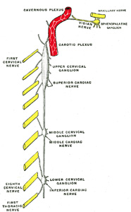
FIG. 844– Diagram of the cervical sympathetic. (Testut.) (See enlarged image)
The Anterior Branches (nn. carotici externi) ramify upon the common carotid artery and upon the external carotid artery and its branches, forming around each a delicate plexus, on the nerves composing which small ganglia are occasionally found. The plexuses accompanying some of these arteries have important communications with other nerves. That surrounding the external maxillary artery communicates with the submaxillary ganglion by a filament; and that accompanying the middle meningeal artery sends an offset to the otic ganglion, and a second, the external petrosal nerve, to the genicular ganglion of the facial nerve.
The middle cervical ganglion (ganglion cervicale medium) is the smallest of the three cervical ganglia, and is occasionally wanting. It is placed opposite the sixth cervical vertebra, usually in front of, or close to, the inferior thyroid artery. It is probably formed by the coalescence of two ganglia corresponding to the fifth and sixth cervical nerves.
The Middle Cardiac Nerve (n. cardiacus medius; great cardiac nerve), the largest of the three cardiac nerves, arises from the middle cervical ganglion, or from the trunk between the middle and inferior ganglia. On the right side it descends behind the common carotid artery, and at the root of the neck runs either in front of or behind the subclavian artery; it then descends on the trachea, receives a few filaments from the recurrent nerve, and joins the right half of the deep part of the cardiac plexus. In the neck, it communicates with the superior cardiac and recurrent nerves. On the left side, the middle cardiac nerve enters the chest between the left carotid and subclavian arteries, and joins the left half of the deep part of the cardiac plexus.
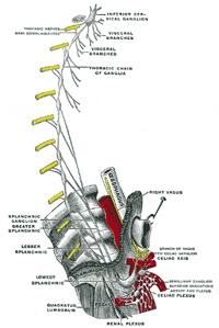
FIG. 845– Plan of right sympathetic cord and splanchnic nerves. (Testut.) (See enlarged image)
The inferior cervical ganglion (ganglion cervicale inferius) is situated between the base of the transverse process of the last cervical vertebra and the neck of the first rib, on the medial side of the costocervical artery. Its form is irregular; it is larger in size than the preceding, and is frequently fused with the first thoracic ganglion. It is probably formed by the coalescence of two ganglia which correspond to the seventh and eighth cervical nerves. It is connected to the middle cervical ganglion by two or more cords, one of which forms a loop around the subclavian artery and supplies offsets to it. This loop is named the ansa subclavia (Vieussenii).
The inferior cardiac nerve (n. cardiacus inferior) arises from either the inferior cervical or the first thoracic ganglion. It descends behind the subclavian artery and along the front of the trachea, to join the deep part of the cardiac plexus. It communicates freely behind the subclavian artery with the recurrent nerve and the middle cardiac nerve.
The offsets to bloodvessels form plexuses on the subclavian artery and its branches. The plexus on the vertebral artery is continued on to the basilar, posterior cerebral, and cerebellar arteries. The plexus on the inferior thyroid artery accompanies the artery to the thyroid gland, and communicates with the recurrent and external laryngeal nerves, with the superior cardiac nerve, and with the plexus on the common carotid artery.
The United States Medical Licensing Examination (USMLE) is a three-step examination for medical licensure in the United States. The Federation of State Medical Boards (FSMB) and the National Board of Medical Examiners (NBME) sponsors USMLE.
The Three Steps of the USMLE
Step 1 tests the important concepts of basic sciences basic to the practice of medicine. It also places special emphasis on principles and mechanisms underlying health, disease, and modes of therapy. Step 1 ensures mastery of the sciences that provide a foundation for the safe and competent practice of medicine. It also tests the scientific principles required for maintenance of competence through lifelong learning.
Step 2 CK tests the medical knowledge, skills, and understanding of clinical science essential for the provision of patient care under supervision. It also includes emphasis on health promotion and disease prevention. Step 2 CK ensures that due attention is devoted to principles of clinical sciences and basic patient-centered skills.
Step 2 CS tests your capacity to practice and provide good medical service in real-life situations. It also tests your communication skills.
Step 3 tests your medical knowledge and understanding of biomedical and clinical science essential for the unsupervised practice of medicine. Step 3 provides a final assessment of physicians assuming independent responsibility for delivering general medical care.