The superficial lymph glands are few and of small size. One or two supratrochlear glands are placed above the medial epicondyle of the humerus, medial to the basilic vein. Their afferents drain the middle, ring, and little fingers, the medial portion of the hand, and the superficial area over the ulnar side of the forearm; these vessels are, however, in free communication with the other lymphatic vessels of the forearm. Their efferents accompany the basilic vein and join the deeper vessels. One or two deltoideopectoral glands are found beside the cephalic vein, between the Pectoralis major and Deltoideus, immediately below the clavicle. They are situated in the course of the external collecting trunks of the arm.
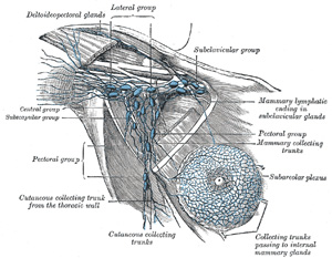
FIG. 607– Lymphatics of the mamma, and the axillary glands (semidiagrammatic). (Poirier and Charpy.) (See enlarged image)
The deep lymph glands are chiefly grouped in the axilla, although a few may be found in the forearm, in the course of the radial, ulnar, and interosseous vessels, and in the arm along the medial side of the brachial artery.
The Axillary Glands (lymphoglandulæ axillares) (Fig. 607) are of large size, vary from twenty to thirty in number, and may be arranged in the following groups:
1. A lateral group of from four to six glands lies in relation to the medial and posterior aspects of the axillary vein; the afferents of these glands drain the whole arm with the exception of that portion whose vessels accompany the cephalic vein. The efferent vessels pass partly to the central and subclavicular groups of axillary glands and partly to the inferior deep cervical glands.
2. An anterior or pectoral group consists of four or five glands along the lower border of the Pectoralis minor, in relation with the lateral thoracic artery. Their afferents drain the skin and muscles of the anterior and lateral thoracic walls, and the central and lateral parts of the namma; their efferents pass partly to the central and partly to the subclavicular groups of axillary glands.
3. A posterior or subscapular group of six or seven glands is placed along the lower margin of the posterior wall of the axilla in the course of the subscapular artery. The afferents of this group drain the skin and muscles of the lower part of the back of the neck and of the posterior thoracic wall; their efferents pass to the central group of axillary glands.
4. A central or intermediate group of three or four large glands is imbedded in the adipose tissue near the base of the axilla. Its afferents are the efferent vessels of all the preceding groups of axillary glands; its efferents pass to the subclavicular group.
5. A medial or subclavicular group of six to twelve glands is situated partly posterior to the upper portion of the Pectoralis minor and partly above the upper border of this muscle. Its only direct territorial afferents are those which accompany the cephalic vein and one which drains the upper peripheral part of the mamma, but it receives the efferents of all the other axillary glands. The efferent vessels of the subclavicular group unite to form the subclavian trunk, which opens either directly into the junction of the internal jugular and subclavian veins or into the jugular lymphatic trunk; on the left side it may end in the thoracic duct. A few efferents from the subclavicular glands usually pass to the inferior deep cervical glands.
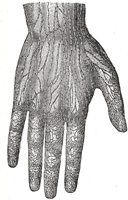
FIG. 608– Lymphatic vessels of the dorsal surface of the hand. (Sappey.) (See enlarged image)
The superficial lymphatic vessels commence (Fig. 608) in the lymphatic plexus which everywhere pervades the skin; the meshes of the plexus are much finer in the palm and on the flexor aspect of the digits than elsewhere. The digital plexuses are drained by a pair of vessels which run on the sides of each digit, and incline backward to reach the dorsum of the hand. From the dense plexus of the palm, vessels pass in different directions, viz., upward toward the wrist, downward to join the digital vessels, medialward to join the vessels on the ulnar border of the hand, and lateralward to those on the thumb. Several vessels from the central part of the plexus unite to form a trunk, which passes around the metacarpal bone of the index finger to join the vessels on the back of that digit and on the back of the thumb. Running upward in front of and behind the wrist, the lymphatic vessels are collected into radial, median, and ulnar groups, which accompany respectively the cephalic, median, and basilic veins in the forearm. A few of the ulnar lymphatics end in the supratrochlear glands, but the majority pass directly to the lateral group of axillary glands. Some of the radial vessels are collected into a trunk which ascends with the cephalic vein to the deltoideopectoral glands; the efferents from this group pass either to the subclavicular axillary glands or to the inferior cervical glands.
The deep lymphatic vessels accompany the deep bloodvessels. In the forearm, they consist of four sets, corresponding with the radial, ulnar, volar, and dorsal interosseous arteries; they communicate at intervals with the superficial lymphatics, and some of them end in the glands which are occasionally found beside the arteries. In their course upward, a few end in the glands which lie upon the brachial artery; but most of them pass to the lateral group of axillary glands.
The United States Medical Licensing Examination (USMLE) is a three-step examination for medical licensure in the United States. The Federation of State Medical Boards (FSMB) and the National Board of Medical Examiners (NBME) sponsors USMLE.
The Three Steps of the USMLE
Step 1 tests the important concepts of basic sciences basic to the practice of medicine. It also places special emphasis on principles and mechanisms underlying health, disease, and modes of therapy. Step 1 ensures mastery of the sciences that provide a foundation for the safe and competent practice of medicine. It also tests the scientific principles required for maintenance of competence through lifelong learning.
Step 2 CK tests the medical knowledge, skills, and understanding of clinical science essential for the provision of patient care under supervision. It also includes emphasis on health promotion and disease prevention. Step 2 CK ensures that due attention is devoted to principles of clinical sciences and basic patient-centered skills.
Step 2 CS tests your capacity to practice and provide good medical service in real-life situations. It also tests your communication skills.
Step 3 tests your medical knowledge and understanding of biomedical and clinical science essential for the unsupervised practice of medicine. Step 3 provides a final assessment of physicians assuming independent responsibility for delivering general medical care.