The veins of the head and neck may be subdivided into three groups: (1) The veins of the exterior of the head and face. (2) The veins of the neck. (3) The diploic veins, the veins of the brain, and the venous sinuses of the dura mater.
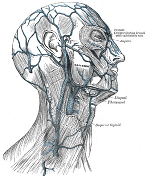
The frontal vein (v. frontalis) begins on the forehead in a venous plexus which communicates with the frontal branches of the superficial temporal vein. The veins converge to form a single trunk, which runs downward near the middle line of the forehead parallel with the vein of the opposite side. The two veins are joined, at the root of the nose, by a transverse branch, called the nasal arch, which receives some small veins from the dorsum of the nose. At the root of the nose the veins diverge, and, each at the medial angle of the orbit, joins the supraorbital vein, to form the angular vein. Occasionally the frontal veins join to form a single trunk, which bifurcates at the root of the nose into the two angular veins.
The supraorbital vein (v. supraorbitalis) begins on the forehead where it communicates with the frontal branch of the superficial temporal vein. It runs downward superficial to the Frontalis muscle, and joins the frontal vein at the medial angle of the orbit to form the angular vein. Previous to its junction with the frontal vein, it sends through the supraorbital notch into the orbit a branch which communicates with the ophthalmic vein; as this vessel passes through the notch, it receives the frontal diploic vein through a foramen at the bottom of the notch.
The angular vein (v. angularis) formed by the junction of the frontal and supraorbital veins, runs obliquely downward, on the side of the root of the nose, to the level of the lower margin of the orbit, where it becomes the anterior facial vein. It receives the veins of the ala nasi, and communicates with the superior ophthalmic vein through the nasofrontal vein, thus establishing an important anastomosis between the anterior facial vein and the cavernous sinus.
The anterior facial vein (v. facialis anterior; facial vein) commences at the side of the root of the nose, and is a direct continuation of the angular vein. It lies behind the external maxillary (facial) artery and follows a less tortuous course. It runs obliquely downward and backward, beneath the Zygomaticus and zygomatic head of the Quadratus labii superioris, descends along the anterior border and then on the superficial surface of the Masseter, crosses over the body of the mandible, and passes obliquely backward, beneath the Platysma and cervical fascia, superficial to the submaxillary gland, the Digastricus and Stylohyoideus. It unites with the posterior facial vein to form the common facial vein, which crosses the external carotid artery and enters the internal jugular vein at a variable point below the hyoid bone. From near its termination a communicating branch often runs down the anterior border of the Sternocleidomastoideus to join the lower part of the anterior jugular vein. The facial vein has no valves, and its walls are not so flaccid as most superficial veins.
Tributaries.—The anterior facial vein receives a branch of considerable size, the deep facial vein, from the pterygoid venous plexus. It is also joined by the superior and inferior palpebral, the superior and inferior labial, the buccinator and the masseteric veins. Below the mandible it receives the submental, palatine, and submaxillary veins, and, generally, the vena comitans of the hypoglossal nerve.
The superficial temporal vein (v. temporalis superficialis) begins on the side and vertex of the skull in a plexus which communicates with the frontal and supraorbital veins, with the corresponding vein of the opposite side, and with the posterior auricular and occipital veins. From this net-work frontal and parietal branches arise, and unite above the zygomatic arch to form the trunk of the vein, which is joined in this situation by the middle temporal vein, from the substance of the Temporalis. It then crosses the posterior root of the zygomatic arch, enters the substance of the parotid gland, and unites with the internal maxillary vein to form the posterior facial vein.
Tributaries.—The superficial temporal vein receives in its course some parotid veins, articular veins from the temporomandibular joint, anterior auricular veins from the auricula, and the transverse facial from the side of the face. The middle temporal vein receives the orbital vein, which is formed by some lateral palpebral branches, and passes backward between the layers of the temporal fascia to join the superficial temporal vein.
The pterygoid plexus (plexus pterygoideus) is of considerable size, and is situated between the Temporalis and Pterygoideus externus, and partly between the two Pterygoidei. It receives tributaries corresponding with the branches of the internal maxillary artery. Thus it receives the sphenopalatine, the middle meningeal, the deep temporal, the pterygoid, masseteric, buccinator, alveolar, and some palatine veins, and a branch which communicates with the ophthalmic vein through the inferior orbital fissure. This plexus communicates freely with the anterior facial vein; it also communicates with the cavernous sinus, by branches through the foramen Vesalii, foramen ovale, and foramen lacerum.
The internal maxillary vein (v. maxillaris interna) is a short trunk which accompanies the first part of the internal maxillary artery. It is formed by a confluence of the veins of the pterygoid plexus, and passes backward between the sphenomandibular ligament and the neck of the mandible, and unites with the temporal vein to form the posterior facial vein.
The posterior facial vein (v. facialis posterior; temporomaxillary vein), formed by the union of the superficial temporal and internal maxillary veins, descends in the substance of the parotid gland, superficial to the external carotid artery but beneath the facial nerve, between the ramus of the mandible and the Sternocleidomastoideus muscle. It divides into two branches, an anterior, which passes forward and unites with the anterior facial vein to form the common facial vein and a posterior, which is joined by the posterior auricular vein and becomes the external jugular vein.
The posterior auricular vein (v. auricularis posterior) begins upon the side of the head, in a plexus which communicates with the tributaries of the occipital, and superficial temporal veins. It descends behind the auricula, and joins the posterior division of the posterior facial vein to form the external jugular. It receive the stylomastoid vein, and some tributaries from the cranial surface of the auricula.
The occipital vein (v. occipitalis) begins in a plexus at the back part of the vertex of the skull, From the plexus emerges a single vessel, which pierces the cranial attachment of the Trapezius and, dipping into the suboccipital triangle, joins the deep cervical and vertebral veins. Occasionally it follows the course of the occipital artery and ends in the internal jugular; in other instances, it joins the posterior auricular and through it opens into the external jugular. The parietal emissary vein connects it with the superior sagittal sinus; and as it passes across the mastoid portion of the temporal bone, it receives the mastoid emissary vein which connects it with the transverse sinus. The occipital diploic vein sometimes joins it.
The United States Medical Licensing Examination (USMLE) is a three-step examination for medical licensure in the United States. The Federation of State Medical Boards (FSMB) and the National Board of Medical Examiners (NBME) sponsors USMLE.
The Three Steps of the USMLE
Step 1 tests the important concepts of basic sciences basic to the practice of medicine. It also places special emphasis on principles and mechanisms underlying health, disease, and modes of therapy. Step 1 ensures mastery of the sciences that provide a foundation for the safe and competent practice of medicine. It also tests the scientific principles required for maintenance of competence through lifelong learning.
Step 2 CK tests the medical knowledge, skills, and understanding of clinical science essential for the provision of patient care under supervision. It also includes emphasis on health promotion and disease prevention. Step 2 CK ensures that due attention is devoted to principles of clinical sciences and basic patient-centered skills.
Step 2 CS tests your capacity to practice and provide good medical service in real-life situations. It also tests your communication skills.
Step 3 tests your medical knowledge and understanding of biomedical and clinical science essential for the unsupervised practice of medicine. Step 3 provides a final assessment of physicians assuming independent responsibility for delivering general medical care.