THE DISTRIBUTION of the systematic arteries is like a highly ramified tree, the common trunk of which, formed by the aorta, commences at the left ventricle, while the smallest ramifications extend to the peripheral parts of the body and the contained organs. Arteries are found in all parts of the body, except in the hairs, nails, epidermis, cartilages, and cornea; the larger trunks usually occupy the most protected situations, running, in the limbs, along the flexor surface, where they are less exposed to injury.
There is considerable variation in the mode of division of the arteries: occasionally a short trunk subdivides into several branches at the same point, as may be observed in the celiac artery and the thyrocervical trunk: the vessel may give off several branches in succession, and still continue as the main trunk, as is seen in the arteries of the limbs; or the division may be dichotomous, as, for instance, when the aorta divides into the two common iliacs.
A branch of an artery is smaller than the trunk from which it arises; but if an artery divides into two branches, the combined sectional area of the two vessels is, in nearly every instance, somewhat greater than that of the trunk; and the combined sectional area of all the arterial branches greatly exceeds that of the aorta; so that the arteries collectively may be regarded as a cone, the apex of which corresponds to the aorta, and the base to the capillary system.
The arteries, in their distribution, communicate with one another, forming what are called anastomoses, and these communications are very free between the large as well as between the smaller branches. The anastomosis between trunks of equal size is found where great activity of the circulation is requisite, as in the brain; here the two vertebral arteries unite to form the basilar, and the two anterior cerebral arteries are connected by a short communicating trunk; it is also found in the abdomen, where the intestinal arteries have very ample anastomoses between their larger branches. In the limbs the anastomoses are most numerous and of largest size around the joints, the branches of an artery above uniting with branches from the vessels below. These anastomoses are of considerable interest to the surgeon, as it is by their enlargement that a collateral circulation is established after the application of a ligature to an artery. The smaller branches of arteries anastomose more frequently than the larger; and between the smallest twigs these anastomoses become so numerous as to constitute a close network that pervades nearly every tissue of the body.
Throughout the body generally the larger arterial branches pursue a fairly straight course, but in certain situations they are tortuous. Thus the external maxillary artery in its course over the face, and the arteries of the lips, are extremely tortuous to accommodate themselves to the movements of the parts. The uterine arteries are also tortuous, to accommodate themselves to the increase of size which the uterus undergoes during pregnancy.
The Pulmonary Artery (A. Pulmonalis) (Figs. 503,504)
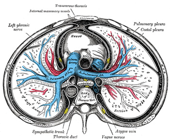
FIG. 503– Transverse section of thorax, showing relations of pulmonary artery. (See enlarged image)
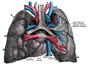
FIG. 504– Pulmonary vessels, seen in a dorsal view of the heart and lungs. The lungs have been pulled away from the median line, and a part of the right lung has been cut away to display the air-ducts and bloodvessels. (See enlarged image)
The pulmonary artery conveys the venous blood from the right ventricle of the heart to the lungs. It is a short, wide vessel, about 5 cm. in length and 3 cm. in diameter, arising from the conus arteriosus of the right ventricle. It extends obliquely upward and backward, passing at first in front and then to the left of the ascending aorta, as far as the under surface of the aortic arch, where it divides, about the level of the fibrocartilage between the fifth and sixth thoracic vertebræ, into right and left branches of nearly equal size.
Relations.—The whole of this vessel is contained within the pericardium. It is enclosed with the ascending aorta in a single tube of the visceral layer of the serous pericardium, which is continued upward upon them from the base of the heart. The fibrous layer of the pericardium is gradually lost upon the external coats of the two branches of the artery. In front, the pulmonary artery is separated from the anterior end of the second left intercostal space by the pleura and left lung, in addition to the pericardium; it rests at first upon the ascending aorta, and higher up lies in front of the left atrium on a plane posterior to the ascending aorta. On either side of its origin is the auricula of the corresponding atrium and a coronary artery, the left coronary artery passing, in the first part of its course, behind the vessel. The superficial part of the cardiac plexus lies above its bifurcation, between it and the arch of the aorta.
The right branch of the pulmonary artery (ramus dexter a. pulmonalis), longer and larger than the left, runs horizontally to the right, behind the ascending aorta and superior vena cava and in front of the right bronchus, to the root of the right lung, where it divides into two branches. The lower and larger of these goes to the middle and lower lobes of the lung; the upper and smaller is distributed to the upper lobe.
The left branch of the pulmonary artery (ramus sinister a. pulmonalis), shorter and somewhat smaller than the right, passes horizontally in front of the descending aorta and left bronchus to the root of the left lung, where it divides into two branches, one for each lobe of the lung.
Above, it is connected to the concavity of the aortic arch by the ligamentum arteriosum, on the left of which is the left recurrent nerve, and on the right the superficial part of the cardiac plexus. Below, it is joined to the upper left pulmonary vein by the ligament of the left vena cava.
The United States Medical Licensing Examination (USMLE) is a three-step examination for medical licensure in the United States. The Federation of State Medical Boards (FSMB) and the National Board of Medical Examiners (NBME) sponsors USMLE.
The Three Steps of the USMLE
Step 1 tests the important concepts of basic sciences basic to the practice of medicine. It also places special emphasis on principles and mechanisms underlying health, disease, and modes of therapy. Step 1 ensures mastery of the sciences that provide a foundation for the safe and competent practice of medicine. It also tests the scientific principles required for maintenance of competence through lifelong learning.
Step 2 CK tests the medical knowledge, skills, and understanding of clinical science essential for the provision of patient care under supervision. It also includes emphasis on health promotion and disease prevention. Step 2 CK ensures that due attention is devoted to principles of clinical sciences and basic patient-centered skills.
Step 2 CS tests your capacity to practice and provide good medical service in real-life situations. It also tests your communication skills.
Step 3 tests your medical knowledge and understanding of biomedical and clinical science essential for the unsupervised practice of medicine. Step 3 provides a final assessment of physicians assuming independent responsibility for delivering general medical care.