Parotideomasseteric Fascia (masseteric fascia).—Covering the Masseter, and firmly connected with it, is a strong layer of fascia derived from the deep cervical fascia. Above, this fascia is attached to the lower border of the zygomatic arch, and behind, it invests the parotid gland.
The Masseter(Fig. 378) is a thick, somewhat quadrilateral muscle, consisting of two portions, superficial and deep. The superficial portion, the larger, arises by a thick, tendinous aponeurosis from the zygomatic process of the maxilla, and from the anterior two-thirds of the lower border of the zygomatic arch; its fibers pass downward and backward, to be inserted into the angle and lower half of the lateral surface of the ramus of the mandible. The deep portion is much smaller, and more muscular in texture; it arises from the posterior third of the lower border and from the whole of the medial surface of the zygomatic arch; its fibers pass downward and forward, to be inserted into the upper half of the ramus and the lateral surface of the coronoid process of the mandible. The deep portion of the muscle is partly concealed, in front, by the superficial portion; behind, it is covered by the parotid gland. The fibers of the two portions are continuous at their insertion.
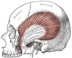
Temporal Fascia.—The temporal fascia covers the Temporalis muscle. It is a strong, fibrous investment, covered, laterally, by the Auricularis anterior and superior, by the galea aponeurotica, and by part of the Orbicularis oculi. The superficial temporal vessels and the auriculotemporal nerve cross it from below upward. Above, it is a single layer, attached to the entire extent of the superior temporal line; but below, where it is fixed to the zygomatic arch, it consists of two layers, one of which is inserted into the lateral, and the other into the medial border of the arch. A small quantity of fat, the orbital branch of the superficial temporal artery, and a filament from the zygomatic branch of the maxillary nerve, are contained between these two layers. It affords attachment by its deep surface to the superficial fibers of the Temporalis.
FIG. 382– The Temporalis; the zygomatic arch and Masseter have been removed. (See enlarged image)
The Temporalis (Temporal muscle) (Fig. 382) is a broad, radiating muscle, situated at the side of the head. It arises from the whole of the temporal fossa (except that portion of it which is formed by the zygomatic bone) and from the deep surface of the temporal fascia. Its fibers converge as they descend, and end in a tendon, which passes deep to the zygomatic arch and is inserted into the medial surface, apex, and anterior border of the coronoid process, and the anterior border of the ramus of the mandible nearly as far forward as the last molar tooth.
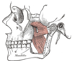
The Pterygoideus externus (External pterygoid muscle) (Fig. 383) is a short, thick muscle, somewhat conical in form, which extends almost horizontally between the infratemporal fossa and the condyle of the mandible. It arises by two heads; an upper from the lower part of the lateral surface of the great wing of the sphenoid and from the infratemporal crest; a lower from the lateral surface of the lateral pterygoid plate. Its fibers pass horizontally backward and lateralward, to be inserted into a depression in front of the neck of the condyle of the mandible, and into the front margin of the articular disk of the temporomandibular articulation.
The Pterygoideus internus (Internal pterygoid muscle) (Fig. 383) is a thick, quadrilateral muscle. It arises from the medial surface of the lateral pterygoid plate and the grooved surface of the pyramidal process of the palatine bone; it has a second slip of origin from the lateral surfaces of the pyramidal process of the palatine and tuberosity of the maxilla. Its fibers pass downward, lateralward, and backward, and are inserted, by a strong tendinous lamina, into the lower and back part of the medial surface of the ramus and angle of the mandible, as high as the mandibular foramen.
Actions.—The Temporalis, Masseter, and Pterygoideus internus raise the mandible against the maxillæ with great force. The Pterygoideus externus assists in opening the mouth, but its main action is to draw forward the condyle and articular disk so that the mandible is protruded and the inferior incisors projected in front of the upper; in this action it is assisted by the Pterygoideus internus. The mandible is retracted by the posterior fibers of the Temporalis. If the Pterygoidei internus and externus of one side act, the corresponding side of the mandible is drawn forward while the opposite condyle remains comparatively fixed, and side-to-side movements. Such as occur during the trituration of food, take place.
The United States Medical Licensing Examination (USMLE) is a three-step examination for medical licensure in the United States. The Federation of State Medical Boards (FSMB) and the National Board of Medical Examiners (NBME) sponsors USMLE.
The Three Steps of the USMLE
Step 1 tests the important concepts of basic sciences basic to the practice of medicine. It also places special emphasis on principles and mechanisms underlying health, disease, and modes of therapy. Step 1 ensures mastery of the sciences that provide a foundation for the safe and competent practice of medicine. It also tests the scientific principles required for maintenance of competence through lifelong learning.
Step 2 CK tests the medical knowledge, skills, and understanding of clinical science essential for the provision of patient care under supervision. It also includes emphasis on health promotion and disease prevention. Step 2 CK ensures that due attention is devoted to principles of clinical sciences and basic patient-centered skills.
Step 2 CS tests your capacity to practice and provide good medical service in real-life situations. It also tests your communication skills.
Step 3 tests your medical knowledge and understanding of biomedical and clinical science essential for the unsupervised practice of medicine. Step 3 provides a final assessment of physicians assuming independent responsibility for delivering general medical care.